Back
Healthcare & Clinical
A Deep Learning Classifier That Separates True-Negative Mammograms from Genuine BIRADS 0 Cases
How Pfactorial Technologies built and benchmarked a CNN pipeline that helps distinguish mammograms unlikely to indicate malignancy from cases genuinely requiring follow-up.

August 21, 2026
Share
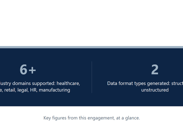
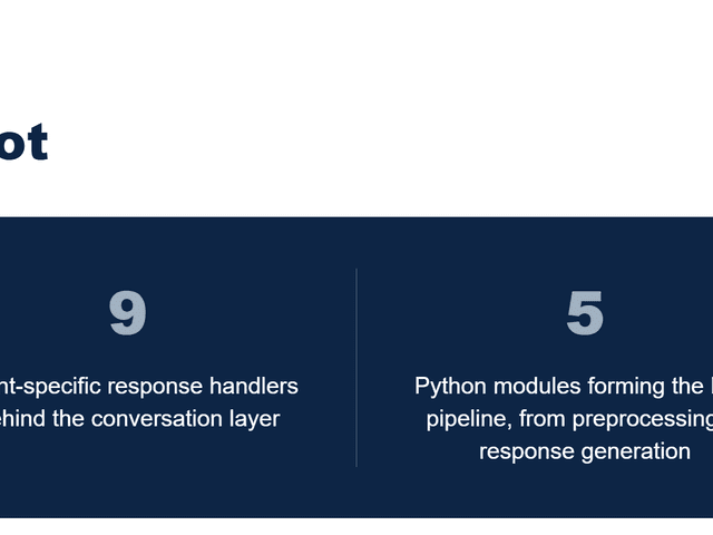
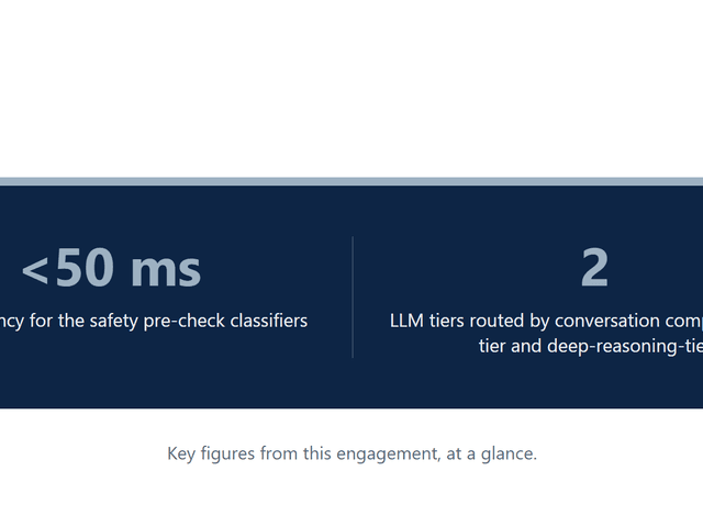
ENGAGEMENT SNAPSHOT

Figure 1 - Key figures from this engagement, at a glance.
EXECUTIVE SUMMARY
Screening mammograms flagged as BIRADS 0 require additional imaging before a radiologist can rule out malignancy, and our client wanted a deep learning classifier that could help distinguish mammograms unlikely to indicate malignancy from genuine cases needing follow-up.
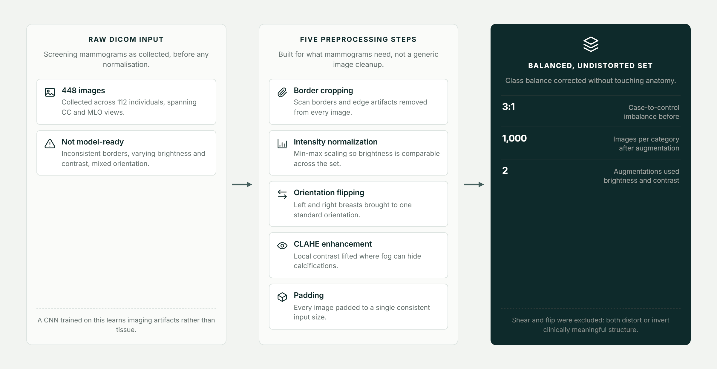
Getting there required more than picking a CNN architecture. Raw mammograms aren't model-ready - inconsistent borders, varying contrast, mixed orientation - and standard image augmentation techniques like flipping can actively corrupt the diagnosis, since breast orientation and tissue structure are themselves clinically meaningful.
Pfactorial built a five-step, diagnostically-aware preprocessing pipeline, a class-balancing strategy that avoided anatomy-distorting augmentation, and benchmarked three training strategies on a ResNet50 architecture - reaching 97% accuracy on the balanced dataset, up from 66% on an unbalanced baseline using the identical architecture.
Why this engagement is representative This engagement shows Pfactorial's ability to diagnose and fix the real bottleneck in a medical imaging model - which was data preprocessing and class balance, not architecture choice - and to prove that with a controlled, benchmarked comparison rather than a single reported number.
THE CHALLENGE
Reaching a usable classifier required solving four problems specific to medical imaging, not general computer vision.
1. Raw mammograms aren't model-ready
Inconsistent borders, varying brightness and contrast, and mixed left/right orientation all have to be normalized before a CNN can learn meaningful patterns rather than imaging artifacts.
2. Standard augmentation can corrupt the diagnosis
Common techniques like flipping or shearing are actively harmful here, since breast orientation and tissue structure are themselves diagnostically meaningful and shouldn't be altered.
3. Class imbalance quietly wrecks accuracy
With roughly three times as many case images as controls, an unbalanced training run learned to guess “case” most of the time rather than genuinely discriminating between categories.
4. A standard CNN wasn't deep enough to find the signal
Mammography features are subtle, and an initial architecture suffered from vanishing gradients, blocking it from reaching useful accuracy.
The real brief Not “train a CNN on mammograms” but “get the preprocessing, augmentation and class balance right first - architecture choice only matters once the data going into it is trustworthy.”
THE SOLUTION
Pfactorial built a domain-aware preprocessing pipeline and a diagnostically-safe augmentation strategy, then isolated variables through controlled training experiments to attribute performance gains to the right cause.

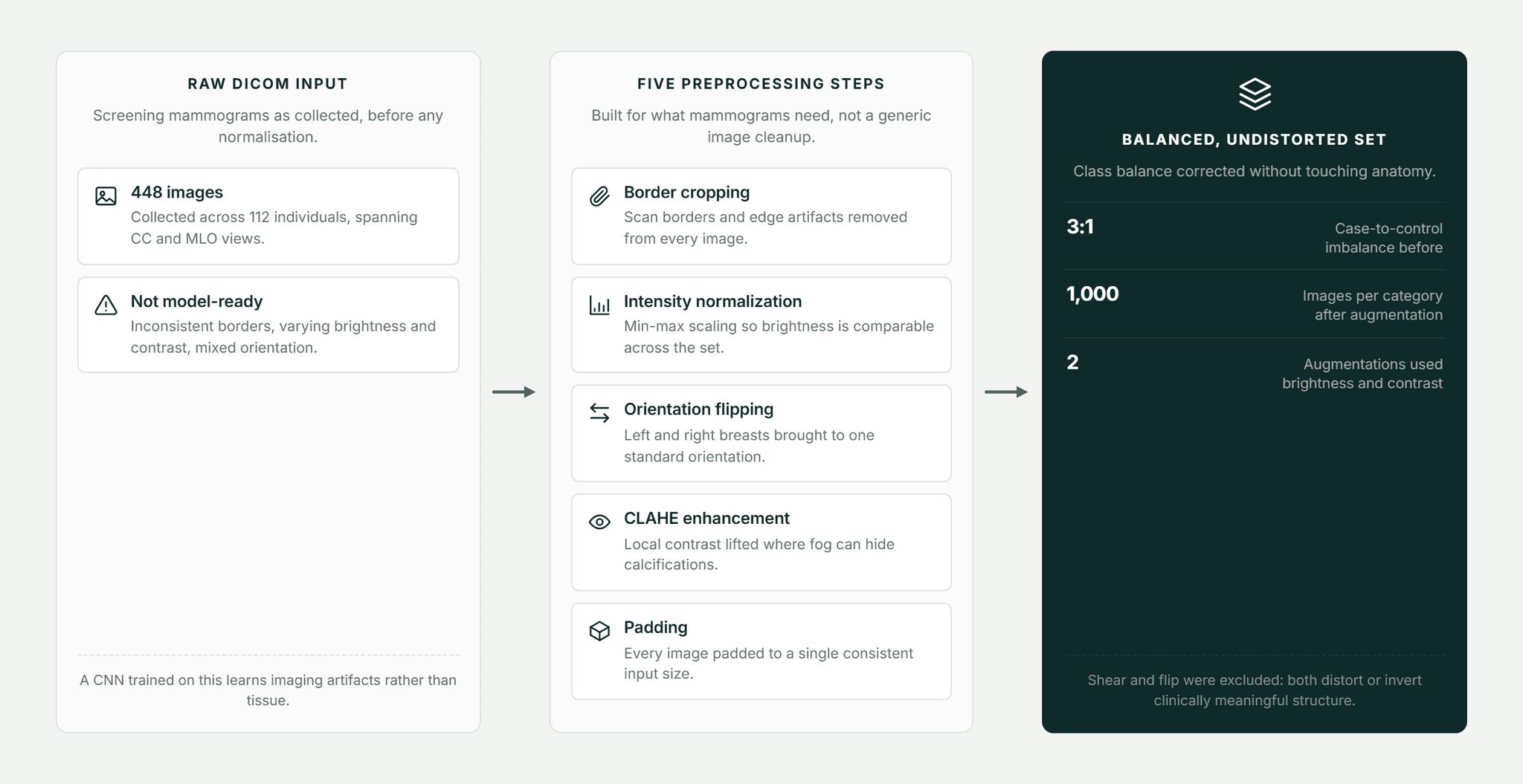
Figure 2 - Five preprocessing steps standardize every image before training begins.
Architectural principles
- Domain-aware preprocessing, not generic cleanup - Border cropping, min-max intensity normalization, orientation standardization, CLAHE contrast enhancement and padding, applied specifically for what mammograms need rather than a generic image-cleaning pass.
- Augmentation that respects diagnostic meaning - Only brightness and contrast variation were used to balance classes; shearing and flipping were deliberately excluded because they would distort or invert clinically relevant structure.
- Architecture chosen to fix a real failure mode - The move from VGG16 to ResNet50 was made specifically because the vanishing-gradient problem was blocking the shallower architecture from learning, not as a default “bigger model” upgrade.
- Isolate variables to know what's actually working - Separate training runs tested unbalanced versus balanced data, and combined versus view-specific (CC-only, MLO-only) datasets, to attribute performance gains to the right cause.
CAPABILITIES DELIVERED
Each deliverable isolates a specific variable's contribution to final model performance.
CAPABILITY | WHAT IT DOES |
|---|---|
5-step preprocessing pipeline | Border cropping, intensity normalization, orientation flipping, CLAHE enhancement, and padding for DICOM images. |
Diagnostically-safe augmentation | Brightness and contrast variation only, fully balancing the case/control dataset. |
Three benchmarked training strategies | Unbalanced baseline, balanced and augmented, and view-specific (CC-only / MLO-only) models. |
ResNet50-based classifier | Selected after VGG16 showed vanishing-gradient limitations on this dataset. |
Full performance reporting | Precision, recall, F1 score and confusion matrices for every training variant. |
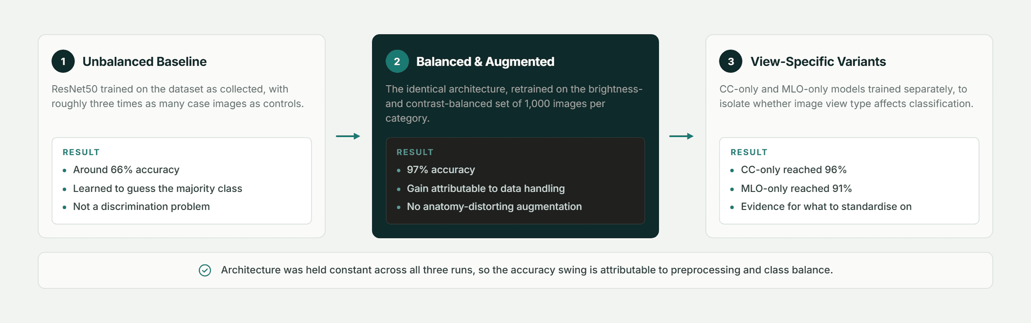
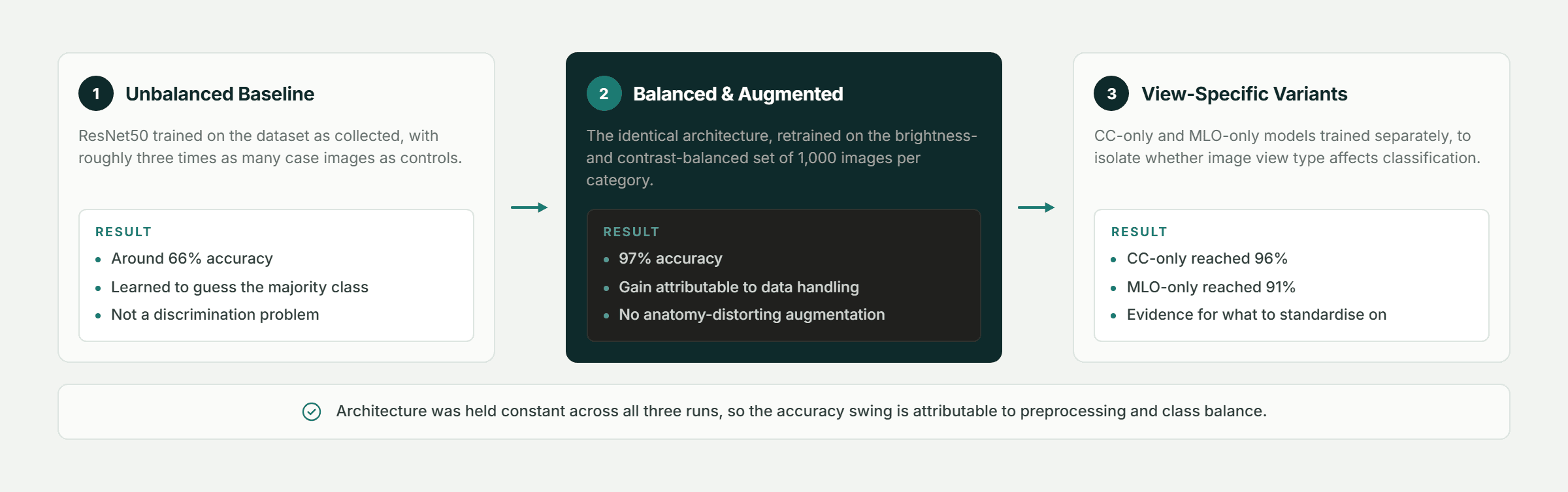
Figure 3 - Same architecture throughout; the accuracy swing comes entirely from data handling.
Design note The 97% accuracy number is meaningless without the 66% unbalanced-dataset run next to it. Same architecture, same images - the difference is entirely in the preprocessing and class balance, which is the actual finding worth remembering.
ENGINEERING FOR SCALE AND RELIABILITY
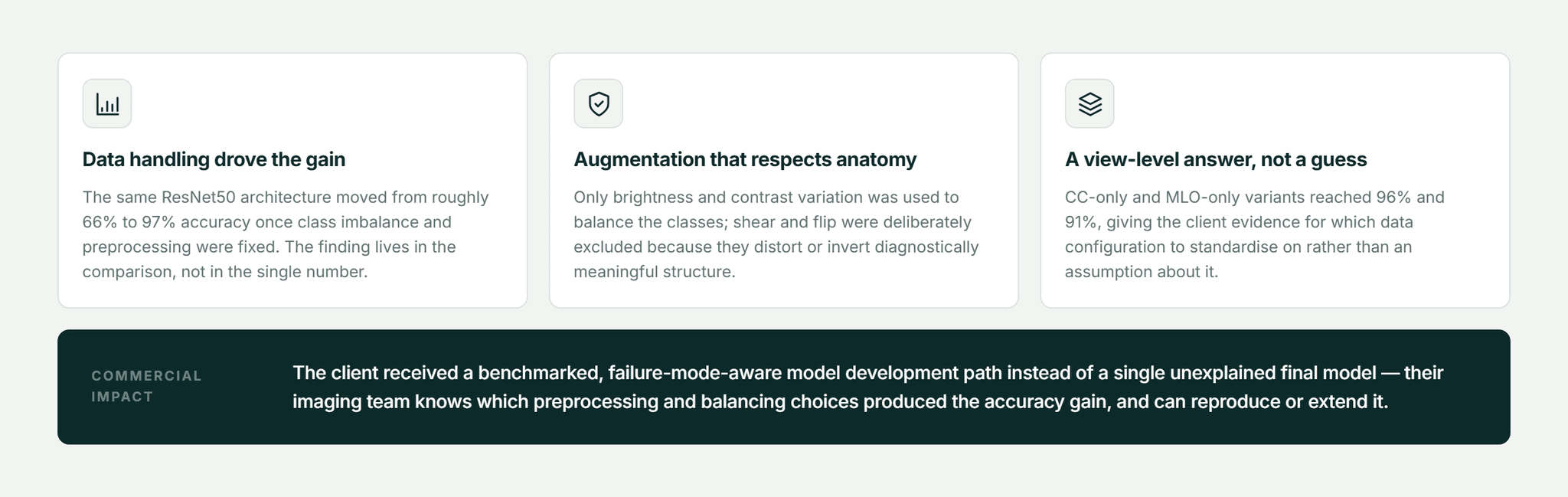
Six methodological decisions ensured the reported accuracy gains reflect real improvement, not an artifact of the evaluation itself.
DICOM-native preprocessing
The pipeline preserves diagnostic orientation and anatomical structure while removing borders, artifacts and inconsistent intensity across the dataset.
Targeted contrast enhancement
CLAHE was applied specifically for foggy or low-clarity images, where subtle findings like calcifications are easiest to miss.
Deliberate augmentation exclusions
Shear and flip augmentation were excluded despite being common elsewhere, because they would distort or invert clinically meaningful anatomical structure.
Class-balanced augmentation
Brightness and contrast augmentation brought the dataset to 1,000 images per category, correcting a roughly 3:1 case-to-control imbalance.
Failure-diagnosed architecture selection
ResNet50 was adopted after diagnosing vanishing gradients as the specific reason VGG16 underperformed, not as a default escalation to a larger model.
View-specific model variants
CC-only and MLO-only models were trained separately to isolate whether image view type affected classification accuracy.
DELIVERY APPROACH
The engagement moved from raw image collection through a controlled, multi-strategy training comparison.
1. Cohort & image collection - 448 mammography images collected across 112 individuals, spanning CC and MLO views.
2. DICOM preprocessing pipeline - border cropping, normalization, orientation flipping, CLAHE enhancement and padding.
3. Diagnostically-safe augmentation strategy - brightness and contrast variation to balance the case/control dataset without distorting anatomy.
4. Baseline model training & failure diagnosis - VGG16 training and identification of the vanishing-gradient limitation.
5. ResNet50 training across 3 strategies - unbalanced baseline, balanced and augmented, and view-specific model variants.
6. Performance benchmarking & reporting - precision, recall, F1 and confusion-matrix reporting for every training strategy.
RESULTS AND IMPACT

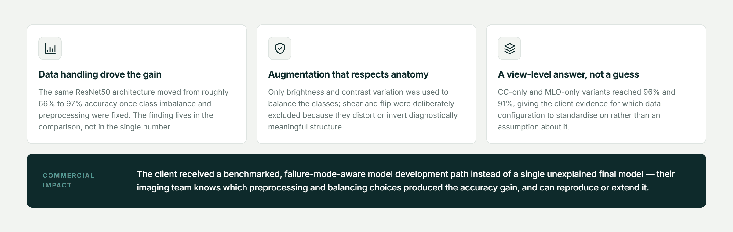
Figure 4 - Key outcomes from this engagement.
The balanced, augmented ResNet50 model reached 97% accuracy, up from roughly 66% on the unbalanced baseline using the identical architecture.
CC-view-only and MLO-view-only variants reached 96% and 91% accuracy respectively, giving the client evidence for which data configuration to standardize on for production use.
What it enabled commercially
The engagement hands the client a benchmarked, failure-mode-aware model development path rather than a single unexplained “final” model, so their imaging or radiology team knows exactly which preprocessing and balancing choices drove the accuracy gain and can reproduce or extend it.
WHY PFACTORIAL
This engagement reflects Pfactorial's medical imaging AI service line: diagnosing the real bottleneck in a computer vision problem - often data handling, not architecture - and proving the fix with a controlled, benchmarked comparison.

Figure 5 - Service lines this engagement draws on.
Engagement enquiries Pfactorial Technologies works with life-sciences and imaging teams that need diagnostic-grade classifiers built with statistical rigor. If you're evaluating a medical imaging AI initiative, we're happy to give you an honest read on scope and risk before anyone commits to anything. · pfactorial.ai

APPENDIX A - TECHNOLOGY STACK
The technology stack underpinning the system, grouped by the layer it serves.

© 2026 Pfactorial Technologies. Client identity and product-specific implementation detail are withheld or generalized; no client data, credentials, source code, or infrastructure detail is included in this document.
Result and Analysis
ENGAGEMENT SNAPSHOT
How Pfactorial Technologies built and benchmarked a CNN pipeline that helps distinguish mammograms unlikely to indicate malignancy from cases genuinely requiring follow-up.

CASE STUDIES

You might also like...

Conversational AI & ChatbotsML Infra, Classifiers & RL
Aug 21, 20268 min readRead

ML Infra, Classifiers & RL
A Machine-Learning Screening Model for Pulmonary Hypertension from Routine Medical Records
Aug 21, 20266 min readRead

Data Scraping & AggregationML Infra, Classifiers & RL
A Risk-Stratification Framework Linking Clinical Milestones to PAH Survival Outcomes
Aug 21, 20266 min readRead

ML Infra, Classifiers & RL
Generating Realistic Synthetic Datasets Without Exposing Real Customer Data
Aug 21, 20268 min readRead

ML Infra, Classifiers & RL
A BERT-Driven Restaurant Chatbot That Classifies Guest Intent, Not Keywords
Aug 21, 20266 min readRead

Automotive & Vehicle
A Browser-Native Neural Network Simulation That Learns to Drive From Experience
Aug 21, 20267 min readRead